


 66 / 64

66
José Manuel Muñoz Olmedo
Basics of isolation and cultivation of chondrocytes
INTRODUCTION
Cartilage tissue is composed of chondrocytes embedded
within a dense extracellular matrix (ECM). It has poor
autonomous regeneration capacity, mainly due to its avascular
nature. Another factor contributing to poor regenerative
capacity of articular cartilage (AC) is the restricted number
of ECM producing cells. The percentage of highly specialized
chondrocytes in cartilage tissue is only 1–3% (1). Chondrocytes
are unable to migrate to a site of injury; they are able to
synthesize fibrous repair tissue, but not sufficiently to fill
even small defects (<3 mm in diameter) with a cartilage-like
matrix, the defects are not repaired and remains permanently
(2). Cartilage was used as one of the first models for research
of
in vitro
engineered tissues and has shown the earliest
application for the cell-based therapy mainly due to its cellular
homogeneity and avascularity (3).
Chondrocytes within their natural environment actively
synthesize and maintain their surrounding matrix. Mature
chondrocytes have limited ability to proliferate; they are often
mislabeled as dormant. In cell culture, human chondrocytes
regain their ability to proliferate. Thus, the basic premise
behind Autologous Chondrocyte Implantation (ACI) is to
overcome the inherent limitations of mature chondrocytes to
effectively restore an injured articular surface.
In monolayer culture, these cells respond by undergoing
rapid proliferation. Histologically, these chondrocytes reversibly
dedifferentiate, assuming a fibroblastic appearance and express
type I collagen as opposed to type II collagen normally seen in
articular cartilage. Once removed from the monolayer culture
and placed in suspension or returned to the articular cartilage
environment, the cells undergo a re-differentiation process
into normal appearing chondrocytes and again produce type II
collagen and proteoglycan aggregates (4).
In 1994 Brittberg
et al
described the use of ACI in treating
full-thickness AC defects of the human knee (5). It was achieved
in a two-stage procedure. Stage 1 involved arthroscopic biopsy
of healthy AC and cultivation of the chondrocytes to produce
between 5 and 10 million cells over a period of 4–6 weeks.
Stage 2 involved debridement of the osteochondral lessions
and coverage by a periosteal flap followed by open implantation
of these cells into the AC defect.
Isolation and cultivation of chondrocytes is a widely spread
technique.ItisrequiredinACIasatreatmentforostheochondritis
dissecans (6), ostheoarthritis (7) and articular cartilage injuries
produced by trauma (8). In general, it can be used in any
articular cartilage injury, where other surgical procedures are
not sufficient (4).
In vitro
chondrocyte manipulation is a crucial
phase of autologous chondrocyte implantation. To minimize
the risk of
in vitro
cell contamination, the manipulation must
be performed in a controlled environment such as a cleanroom
according to good laboratory practice (GLP). The GLP standards
provide guidance on implementing GLP requirements critical
for laboratory operations (9, 10).
In the current study, we have used basic methods
for isolation and cultivation of chondrocytes from human
articular cartilage according to GLP standards. Phenotype
characterization of chondrocytes was performed by flow
cytometry analysis.
MATERIALS AND METHODS
Work in cleanroom according to GLP standards
Isolation and cultivation of chondrocytes was performed
in the cleanroom in Associated Tissue Bank of Faculty of
Medicine of P. J. Šafárik University and L. Pasteur University
Hospital in Košice, Slovakia. A cleanroom is a controlled
environment in which the concentration of airborne particles is
controlled to specified limits (Tab.1). Contaminants generated
by people, process, facilities and equipment were continually
removed from the air. Air flow rates, direction, pressurization,
temperature, humidity and specialized filtration were all
tightly controlled. Persons and materials entered and exited
the cleanroom through airlocks (material pass box), gowning
rooms and they weared special clothing designed to trap
contaminants that are naturally generated by skin and the
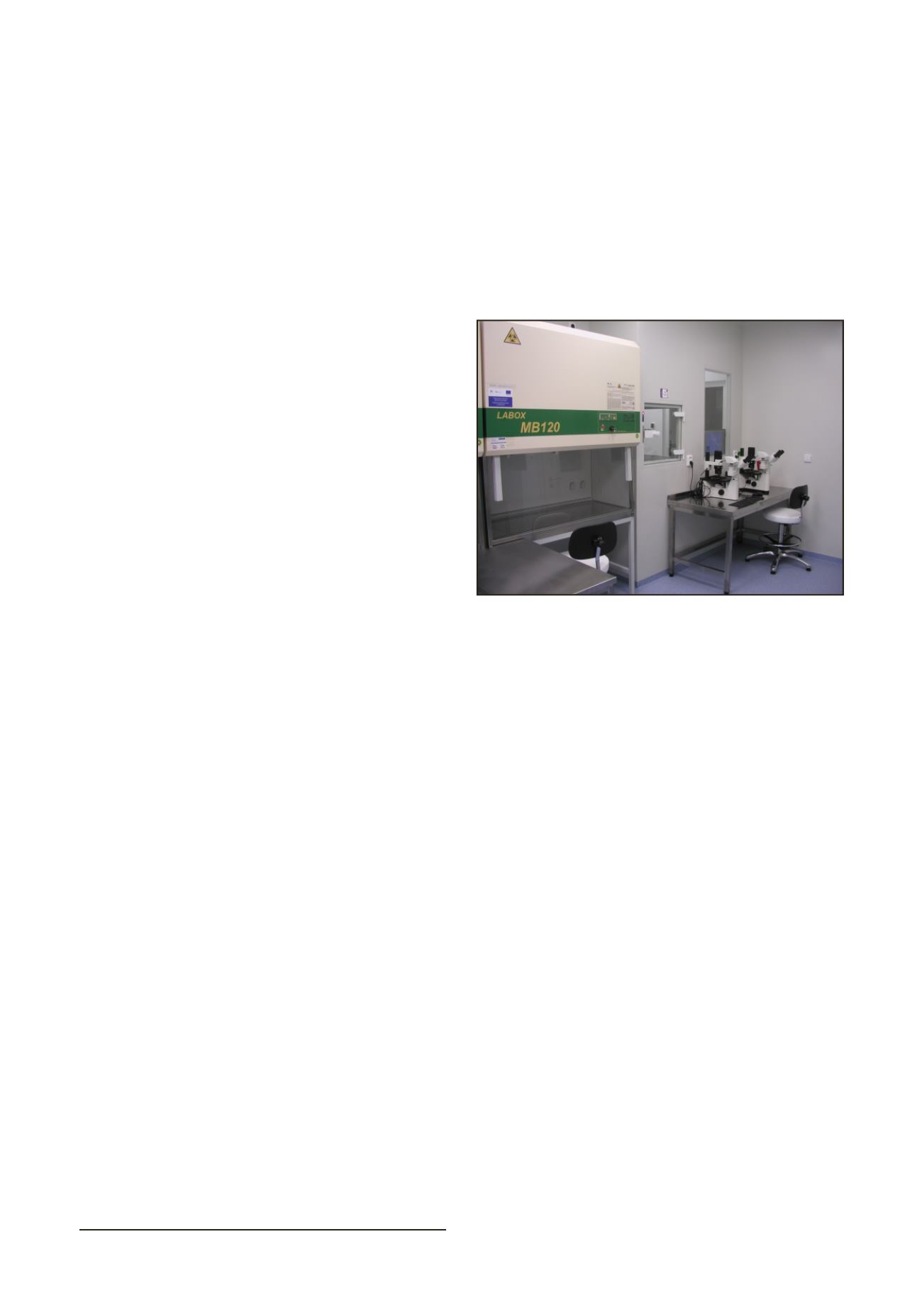
body. Cells were handled inside biological safety cabinets and
cultivated in the gas incubators (Fig. 1).
Isolation and cultivation of chondrocytes
Human cartilage tissue was harvested from the lateral
femoral condyle of 5 patients (an average age: 63 years)
undergoing total knee replacement surgery due to osteoarthritis
(stage 3). Cartilage tissue was harvested in accordance with the
ethical standards of L. Pasteur University Hospital commitee
on human experimentation in Košice, Slovakia. Cartilage
tissue was placed into the transport medium containing sterile
high-glucose Dulbecco’s modified Eagle medium (DMEM;
Invitrogen, GIBCO, USA) supplemented with 1% antibiotic/
antimycotic solution (10,000 units/mL penicillin, 10,000
µ
g/mL
streptomycin, and 25
µ
g/mL amphotericin B; Invitrogen, GIBCO,
USA). Cartilage tissue was minced with a scalpel to small pieces
(1x1x1 mm) and digested with 0,1% bacterial collagenase type
II (Invitrogen, GIBCO, USA) in Ham’s F-12 (Biochrom AG) for
20 h at 37 C in 95% air and 5% CO
2
humified atmosphere. Cell
suspension was filtered by 40
µ
m nylon cell strainer (BD Falcon,
Biosciences, Bedford, MA) to remove cell raft and matrix debris.
The filtrate was then centrifuged at 150 x g for 7 min and the
pellet washed twice with DMEM (Invitrogen, GIBCO, USA).
Isolated cells were suspended in cell culture medium containing
Ham’s F-12 (Biochrom AG), 10% fetal bovine serum (FBS;
Invitrogen, GIBCO, USA), 1% antibiotic/antimycotic solution
(10,000 units/mL penicillin, 10,000
µ
g/mL streptomycin, and 25
µ
g/mL amphotericin B; Invitrogen, GIBCO, USA) and 1% Insulin-
Transferin-Selenium–A supplement (Invitrogen, GIBCO, USA).
Chondrocytes were cultivated as a monolayer for expansion in
37 C humidified incubator with an atmosphere of 95% air and
5% CO
2
. The medium was changed 2 times weekly. Confluent
layers of chondrocytes were dissociated with 0.05% Trypsin-
EDTA solution (Invitrogen, GIBCO®, USA) and the number
and viability of cells was assessed by TC10™ Automated Cell
Counter (Bio-Rad Laboratories).
Characterization of chondrocytes
Fig. 1.
Cleanroom facilities for cell cultivation according to GLP
standards